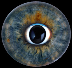
Case 15Roth Spots / Bacterial Endocarditis
Clinical Presentation
A 12-year-old girl enters the hospital for evaluation of fever and generalized weakness. She states her vision suddenly became blurred this week. Both eyes have similar findings.
Learner Questions
- Describe the ophthalmoscopic findings.
- What are the histopathologic correlates of these findings?
- What questions should be asked in the review of systems? What physical findings would you look for?
- What is the most likely diagnosis?
Answer Framework
Findings
ROTH SPOTS - flame/boat-shaped intraretinal hemorrhages with WHITE or PALE CENTERS (fibrin-platelet thrombus from septic embolus + leukocytic infiltrate). Distributed around the posterior pole and disc. Disc edema may be present. Minimal vitreous reaction.
ROS + exam
ROS: congenital heart disease, prior dental work/infections, joint pain, SOB. Physical (Duke Criteria): new/changed cardiac murmur, Osler nodes (tender finger/toe nodules - immune complex), Janeway lesions (non-tender palmar/plantar macules - septic emboli), splinter hemorrhages, petechiae, splenomegaly.
Histopathology
Septic microemboli lodge in retinal capillaries -> fibrin-platelet thrombus + inflammatory response -> capillary wall disruption -> surrounding hemorrhage. White center = fibrin thrombus + lymphocytes/macrophages. Risk of endophthalmitis if emboli seed the vitreous cavity.
Diagnosis
Infective Endocarditis (SBE) on congenital heart disease - Strep viridans most common at this age. Workup: Blood cultures x 3 (separate sites BEFORE antibiotics - most important test), TTE/TEE echocardiography (vegetations = major Duke criterion), CBC, ESR/CRP, urinalysis (microscopic hematuria from immune complex glomerulonephritis).
Teaching Pearl
Roth spots = white-centered retinal hemorrhages = intraretinal septic emboli -> demand blood cultures and echocardiography in any febrile patient. In a child, suspect underlying congenital heart disease. The peripheral stigmata of IE (Janeway, Osler, splinter hemorrhages, Roth spots) all represent the same pathophysiology: sustained bacteremia -> septic emboli + immune complex deposition.
Original answer transcript
Findings ROS + exam
ROTH SPOTS - flame/boat-shaped intraretinal hemorrhages with WHITE or PALE CENTERS ROS: congenital heart disease, prior dental work/infections, joint pain, SOB. Physical (Duke
(fibrin-platelet thrombus from septic embolus + leukocytic infiltrate). Distributed around Criteria): new/changed cardiac murmur, Osler nodes (tender finger/toe nodules - immune
the posterior pole and disc. Disc edema may be present. Minimal vitreous reaction. complex), Janeway lesions (non-tender palmar/plantar macules - septic emboli), splinter
hemorrhages, petechiae, splenomegaly.
Histopathology Diagnosis
Septic microemboli lodge in retinal capillaries -> fibrin-platelet thrombus + inflammatory Infective Endocarditis (SBE) on congenital heart disease - Strep viridans most common at
response -> capillary wall disruption -> surrounding hemorrhage. White center = fibrin this age. Workup: Blood cultures x 3 (separate sites BEFORE antibiotics - most important
thrombus + lymphocytes/macrophages. Risk of endophthalmitis if emboli seed the vitreous test), TTE/TEE echocardiography (vegetations = major Duke criterion), CBC, ESR/CRP,
cavity. urinalysis (microscopic hematuria from immune complex glomerulonephritis).
TEACHING PEARL
Roth spots = white-centered retinal hemorrhages = intraretinal septic emboli -> demand blood cultures and echocardiography in any febrile patient. In a child, suspect
underlying congenital heart disease. The peripheral stigmata of IE (Janeway, Osler, splinter hemorrhages, Roth spots) all represent the same pathophysiology: sustained
bacteremia -> septic emboli + immune complex deposition.