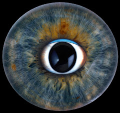
Pupillary response
RAPD (Marcus Gunn pupil) in the affected eye. Swinging flashlight test: both pupils paradoxical the fellow eye. RAPD = asymmetric optic nerve or extensive retinal lesion.
Case 03 - Neuro-ophthalmology, orbit, and inflammation
42-year-old African-American woman - Painless monocular vision loss progressing over months
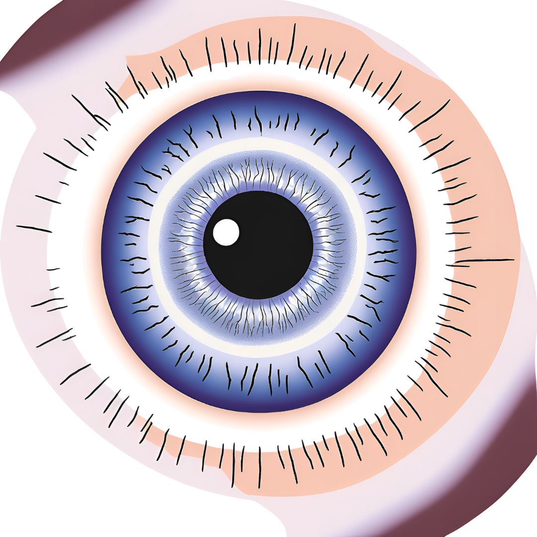
A 42-year-old African-American woman complains of painless loss of vision in one eye which has progressed during the past several months. The optic nerve in the affected eye is illustrated. The opposite eye is normal.
RAPD (Marcus Gunn pupil) in the affected eye. Swinging flashlight test: both pupils paradoxical the fellow eye. RAPD = asymmetric optic nerve or extensive retinal lesion.
Pale, swollen disc with edema and pallor - infiltrative or compressive optic neuropathy. In thi the leading diagnosis.
Inflammatory (sarcoidosis #1 in this demographic, MS, syphilis, TB, Lyme) - Compressive (orbital mass, meningioma, pituitary) - Infiltrative (lymphoma, sarcoid) - Ischemic (NAION) - Glaucomatous (NTG) - Hereditary (Leber's LHON). Work-up: ACE, CXR/CT, RPR, PPD, biopsy.
Sarcoidosis predilects young African-American women. Ocular involvement in ~25-50% of cases; optic nerve granulomas are vision-threatening. Work-up: ACE, serum lysozyme, chest CT (hilar adenopathy), RPR (always rule out syphilis). Treatment: systemic corticosteroids.
Pupillary response
RAPD (Marcus Gunn pupil) in the affected eye. Swinging flashlight test: both pupils paradoxically dilate when light swings to the affected eye (weaker afferent signal). Normal direct/consensual in
the fellow eye. RAPD = asymmetric optic nerve or extensive retinal lesion.
Optic disc
Pale, swollen disc with edema and pallor - infiltrative or compressive optic neuropathy. In this patient (African-American woman, painless, gradual): sarcoid granuloma infiltrating the optic nerve is
the leading diagnosis.
DDx
Inflammatory (sarcoidosis #1 in this demographic, MS, syphilis, TB, Lyme) - Compressive (orbital mass, meningioma, pituitary) - Infiltrative (lymphoma, sarcoid) - Ischemic (NAION) - Glaucomatous
(NTG) - Hereditary (Leber's LHON). Work-up: ACE, CXR/CT, RPR, PPD, biopsy.
TEACHING PEARL
Sarcoidosis predilects young African-American women. Ocular involvement in ~25-50% of cases; optic nerve granulomas are vision-threatening. Work-up: ACE, serum
lysozyme, chest CT (hilar adenopathy), RPR (always rule out syphilis). Treatment: systemic corticosteroids.