Case 10Ocular Sarcoidosis / Uveitis
Clinical Presentation
A 35-year-old African-American woman is admitted for evaluation of fever of unknown origin. She recalls several episodes of bilateral photophobia, decreased vision, and mild discomfort. Signs are asymmetric in both eyes.
Learner Questions
- Explain this patient's ocular symptoms.
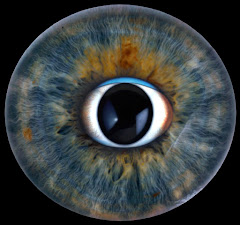
- Describe and explain the ocular findings shown in the photograph.
- What is the differential diagnosis? What laboratory tests would you need?
- How should the ocular manifestations be treated?
Answer Framework
Symptoms
Bilateral photophobia = ciliary muscle spasm from iridocyclitis (ciliary body inflammation -> pain with light and accommodation). Decreased vision = cells + flare in AC, corneal edema from KPs, cystoid macular edema (chronic uveitis). Mild discomfort = ciliary spasm + mild IOP elevation.
DDx + workup
DDx: Sarcoidosis (#1 in this demographic), Syphilis (ALWAYS TEST FIRST - the great masquerader), TB, VKH (Asian/Hispanic, meningismus, vitiligo/poliosis), Sympathetic ophthalmia. Labs: RPR+FTA-ABS, ACE, serum lysozyme, CXR/CT chest (hilar adenopathy), QuantiFERON-Gold, ANA, gallium scan ('panda sign').
Findings
MUTTON-FAT KPs - large, greasy, yellowish-white aggregates of macrophages and epithelioid cells on inferior corneal endothelium. PATHOGNOMONIC for GRANULOMATOUS uveitis. Smaller KPs superiorly. Posterior synechiae (iris-lens adhesions) may be present.
Treatment
Topical prednisolone acetate 1% (q1h initially -> taper) + cycloplegic (prevents posterior synechiae). Sub-Tenon's or intravitreal triamcinolone for refractory CME. Systemic steroids for bilateral severe/posterior disease. Steroid-sparing: methotrexate or mycophenolate for chronic disease. Monitor IOP throughout.
Teaching Pearl
Uveitis mantra: ALWAYS rule out syphilis first. Mutton-fat KPs = GRANULOMATOUS uveitis. Granulomatous causes (SSTVLG): Sarcoid, Syphilis, TB, VKH, Leprosy, Granulomatosis with polyangiitis. Recurrent bilateral granulomatous uveitis in a young Black woman = sarcoidosis until proven otherwise.
Original answer transcript
Symptoms DDx + workup
Bilateral photophobia = ciliary muscle spasm from iridocyclitis (ciliary body inflammation -> DDx: Sarcoidosis (#1 in this demographic), Syphilis (ALWAYS TEST FIRST - the great
pain with light and accommodation). Decreased vision = cells + flare in AC, corneal edema masquerader), TB, VKH (Asian/Hispanic, meningismus, vitiligo/poliosis), Sympathetic
from KPs, cystoid macular edema (chronic uveitis). Mild discomfort = ciliary spasm + mild ophthalmia. Labs: RPR+FTA-ABS, ACE, serum lysozyme, CXR/CT chest (hilar adenopathy),
IOP elevation. QuantiFERON-Gold, ANA, gallium scan ('panda sign').
Findings Treatment
MUTTON-FAT KPs - large, greasy, yellowish-white aggregates of macrophages and Topical prednisolone acetate 1% (q1h initially -> taper) + cycloplegic (prevents posterior
epithelioid cells on inferior corneal endothelium. PATHOGNOMONIC for GRANULOMATOUS synechiae). Sub-Tenon's or intravitreal triamcinolone for refractory CME. Systemic steroids
uveitis. Smaller KPs superiorly. Posterior synechiae (iris-lens adhesions) may be present. for bilateral severe/posterior disease. Steroid-sparing: methotrexate or mycophenolate for
chronic disease. Monitor IOP throughout.
TEACHING PEARL
Uveitis mantra: ALWAYS rule out syphilis first. Mutton-fat KPs = GRANULOMATOUS uveitis. Granulomatous causes (SSTVLG): Sarcoid, Syphilis, TB, VKH, Leprosy,
Granulomatosis with polyangiitis. Recurrent bilateral granulomatous uveitis in a young Black woman = sarcoidosis until proven otherwise.