Findings
Dot-and-blot hemorrhages, hard exudates (yellow-white lipid deposits, sharp borders), and microaneurysms bilaterally. No neovascularization - consistent with NPDR.
Case 01 - Retina and vascular pathology
60-year-old obese woman - General malaise - No ocular complaints - Bilateral fundus changes
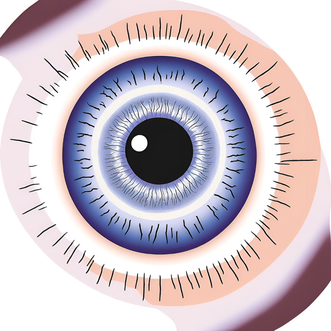
A 60-year-old obese woman complains of general malaise and has no ocular complaints. The fundus photograph illustrates the ophthalmoscopic findings in both eyes.
Dot-and-blot hemorrhages, hard exudates (yellow-white lipid deposits, sharp borders), and microaneurysms bilaterally. No neovascularization - consistent with NPDR.
Duration & type of DM, HbA1c, BP, renal function (nephropathy correlates with severity), lipid levels, prior laser, medications, family history of DM complications.
Microaneurysms = pericyte loss -> capillary outpouching (earliest change). Hard exudates = inner BRB breakdown -> lipoproteins in outer plexiform/Henle's layer. Blot hemorrhages = deeper retinal layer capillary rupture.
Non-Proliferative Diabetic Retinopathy (NPDR). DDx: Hypertensive retinopathy, HIV retinopathy, CRVO. Severity by ETDRS '4-2-1 rule': hemorrhages in 4 quadrants, venous beading >=2 quadrants, or IRMA >=1 quadrant = severe NPDR.
DR is the leading cause of new blindness in working-age adults in the developed world. Tight glycemic control (DCCT/UKPDS) and annual dilated exams are the cornerstones of prevention. Refer to retina when CSME is present - edema within 500 um of the foveal center.
Findings Key history
Dot-and-blot hemorrhages, hard exudates (yellow-white lipid deposits, sharp borders), and Duration & type of DM, HbA1c, BP, renal function (nephropathy correlates with severity),
microaneurysms bilaterally. No neovascularization - consistent with NPDR. lipid levels, prior laser, medications, family history of DM complications.
Histopathology Diagnosis
Microaneurysms = pericyte loss -> capillary outpouching (earliest change). Hard exudates = Non-Proliferative Diabetic Retinopathy (NPDR). DDx: Hypertensive retinopathy, HIV
inner BRB breakdown -> lipoproteins in outer plexiform/Henle's layer. Blot hemorrhages = retinopathy, CRVO. Severity by ETDRS '4-2-1 rule': hemorrhages in 4 quadrants, venous
deeper retinal layer capillary rupture. beading >=2 quadrants, or IRMA >=1 quadrant = severe NPDR.
TEACHING PEARL
DR is the leading cause of new blindness in working-age adults in the developed world. Tight glycemic control (DCCT/UKPDS) and annual dilated exams are the
cornerstones of prevention. Refer to retina when CSME is present - edema within 500 um of the foveal center.