Case 11Giant Cell Arteritis / AION
Clinical Presentation
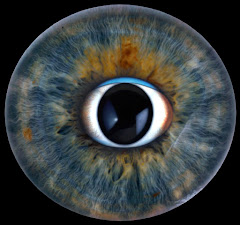
A 70-year-old woman suffered sudden but painless vision loss in one eye. She has also experienced headaches and shoulder pain for the past several months. The affected eye is shown; her other eye remains normal.
Learner Questions
- What questions would you include when taking the history?
- Describe the ophthalmoscopic findings.
- What is the differential diagnosis?
- What should the diagnostic workup include?
- What are your strategies for short- and long-term management?
Answer Framework
History
Jaw claudication (most specific - ~65% sensitive), scalp tenderness, temporal artery tenderness/nodularity, PMR symptoms (proximal girdle stiffness worse in AM), prior transient monocular VL, constitutional symptoms (fever, weight loss), diplopia, prior elevated ESR/CRP.
Workup
START STEROIDS IMMEDIATELY - do not wait for biopsy results. ESR (Westergren, often >100) + CRP (more sensitive - elevated in nearly all active GCA). CBC (thrombocytosis, anemia). Temporal artery biopsy (>=2 cm, within 1-2 weeks after starting steroids - histology remains positive). Color duplex US (halo sign around temporal arteries). PET-CT for large-vessel involvement.
Findings
Pale, swollen optic disc with CHALKY-WHITE PALLOR (more profound than NAION) and blurred margins. Possible segmental/altitudinal disc edema. Flame hemorrhages at disc margin. Chalky-white disc = hallmark of arteritic AION. Concurrent cilioretinal artery occlusion possible.
Management
Acute: IV methylprednisolone 250-500 mg q6h if vision already lost (may protect fellow eye). Oral prednisone 1 mg/kg/day if vision intact. Long-term: slow taper over 1-2 years guided by ESR/CRP. Tocilizumab (IL-6 inhibitor - GiACTA trial) = steroid-sparer, reduces relapse + cumulative steroid dose. Low-dose aspirin 81 mg. Annual CXR for aortic aneurysm.
DDx
Arteritic AION (GCA) - top here: chalky disc + systemic symptoms + elevated ESR/CRP. Non-arteritic AION (NAION) - small disc-at-risk, vasculopathic risk factors (HTN/DM/sleep apnea), normal inflammatory markers, altitudinal field defect. CRAO - cherry-red spot. PION - normal disc acutely. Optic neuritis - younger, painful.
Teaching Pearl
GCA is the ONLY vasculitis causing blindness - a true ophthalmic emergency. Risk to fellow eye = 25-50% within days WITHOUT treatment. Treat with steroids IMMEDIATELY - biopsy can wait 1-2 weeks (histology remains positive). Tocilizumab (IL-6 inhibitor, GiACTA trial) is the first approved biologic for GCA. Vision lost to arteritic AION rarely recovers (<15%).
Original answer transcript
History Workup
START STEROIDS IMMEDIATELY - do not wait for biopsy results. ESR (Westergren, often
Jaw claudication (most specific - ~65% sensitive), scalp tenderness, temporal artery >100) + CRP (more sensitive - elevated in nearly all active GCA). CBC (thrombocytosis,
tenderness/nodularity, PMR symptoms (proximal girdle stiffness worse in AM), prior anemia). Temporal artery biopsy (>=2 cm, within 1-2 weeks after starting steroids -
transient monocular VL, constitutional symptoms (fever, weight loss), diplopia, prior histology remains positive). Color duplex US (halo sign around temporal arteries). PET-CT
elevated ESR/CRP. for large-vessel involvement.
Findings Management
Acute: IV methylprednisolone 250-500 mg q6h if vision already lost (may protect fellow
Pale, swollen optic disc with CHALKY-WHITE PALLOR (more profound than NAION) and
eye). Oral prednisone 1 mg/kg/day if vision intact. Long-term: slow taper over 1-2 years
blurred margins. Possible segmental/altitudinal disc edema. Flame hemorrhages at disc guided by ESR/CRP. Tocilizumab (IL-6 inhibitor - GiACTA trial) = steroid-sparer, reduces
margin. Chalky-white disc = hallmark of arteritic AION. Concurrent cilioretinal artery relapse + cumulative steroid dose. Low-dose aspirin 81 mg. Annual CXR for aortic
occlusion possible.
aneurysm.
DDx
Arteritic AION (GCA) - top here: chalky disc + systemic symptoms + elevated ESR/CRP.
Non-arteritic AION (NAION) - small disc-at-risk, vasculopathic risk factors (HTN/DM/sleep
apnea), normal inflammatory markers, altitudinal field defect. CRAO - cherry-red spot.
PION - normal disc acutely. Optic neuritis - younger, painful.
TEACHING PEARL
GCA is the ONLY vasculitis causing blindness - a true ophthalmic emergency. Risk to fellow eye = 25-50% within days WITHOUT treatment. Treat with steroids
IMMEDIATELY - biopsy can wait 1-2 weeks (histology remains positive). Tocilizumab (IL-6 inhibitor, GiACTA trial) is the first approved biologic for GCA. Vision lost to
arteritic AION rarely recovers (<15%).