Case 07Choroidal Melanoma
Clinical Presentation
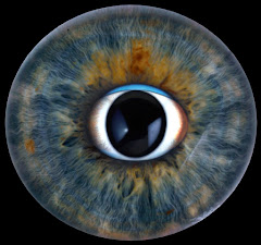
This 55-year-old white man comes to your office with complaints of painless but progressive visual field loss in one eye. The accompanying photograph was taken from the affected eye.
Learner Questions
- When evaluating the patient's history, what questions would you ask?
- What diagnostic tests would you order?
- What would be your differential diagnosis?
- What treatment options would you consider?
Answer Framework
History
Prior cutaneous melanoma, family history (BAP1 syndrome, uveal melanoma), choroidal nevi, duration/progression, floaters/flashes (associated RD), weight loss/abdominal pain/jaundice (liver metastases - preferred metastatic site), prior ocular trauma/radiation.
Diagnostics
B-scan US (acoustic hollowness, choroidal excavation, orbital shadowing - measures tumor height; key test). MRI orbits (T1 hyperintense, T2 hypointense - melanin). Systemic staging: LFTs, LDH, liver MRI/US, CXR. Genetic testing: monosomy 3 + 8q amplification = high metastatic risk.
DDx
Choroidal melanoma (most common primary intraocular malignancy in adults), choroidal metastasis (breast #1 in women, lung #1 in men - overall most common intraocular malignancy), choroidal hemangioma (orange-red, posterior pole), choroidal nevus (flat, <2 mm, no SRF - ABCDE risk factors), disciform scar (AMD). Treatment Small: observation vs. TTT/plaque brachytherapy. Medium: I-125 plaque brachytherapy (COMS trial gold standard). Large: enucleation. Proton beam for posterior tumors.
Teaching Pearl
Choroidal melanoma has 50% 10-year metastatic rate regardless of treatment - liver MRI every 6-12 months for life is mandatory. Monosomy 3 on cytogenetics = highest metastatic risk. ABCDE nevus risk factors for transformation: subretinal fluid, symptoms, Diameter >5 mm, Edge touchni g disc, Orange lipofuscin pigment.
Original answer transcript
History
Prior cutaneous melanoma, family history (BAP1 syndrome, uveal melanoma), choroidal
nevi, duration/progression, floaters/flashes (associated RD), weight loss/abdominal
pain/jaundice (liver metastases - preferred metastatic site), prior ocular trauma/radiation.
Diagnostics
B-scan US (acoustic hollowness, choroidal excavation, orbital shadowing - measures
tumor height; key test). MRI orbits (T1 hyperintense, T2 hypointense - melanin). Systemic
staging: LFTs, LDH, liver MRI/US, CXR. Genetic testing: monosomy 3 + 8q amplification =
high metastatic risk.
DDx
Choroidal melanoma (most common primary intraocular malignancy in adults), choroidal
metastasis (breast #1 in women, lung #1 in men - overall most common intraocular
malignancy), choroidal hemangioma (orange-red, posterior pole), choroidal nevus (flat, <2
mm, no SRF - ABCDE risk factors), disciform scar (AMD).
Treatment
Small: observation vs. TTT/plaque brachytherapy. Medium: I-125 plaque brachytherapy
(COMS trial gold standard). Large: enucleation. Proton beam for posterior tumors.
Metastatic: liver-directed therapy. NOTE: uveal melanoma does NOT respond to
ipilimumab/checkpoint inhibitors (GNA11/GNAQ mutations, not BRAF).
TEACHING PEARL
Choroidal melanoma has 50% 10-year metastatic rate regardless of treatment - liver MRI every 6-12 months for life is mandatory. Monosomy 3 on cytogenetics =
highest metastatic risk. ABCDE nevus risk factors for transformation: subretinal fluid, symptoms, Diameter >5 mm, Edge touchni g disc, Orange lipofuscin pigment.